
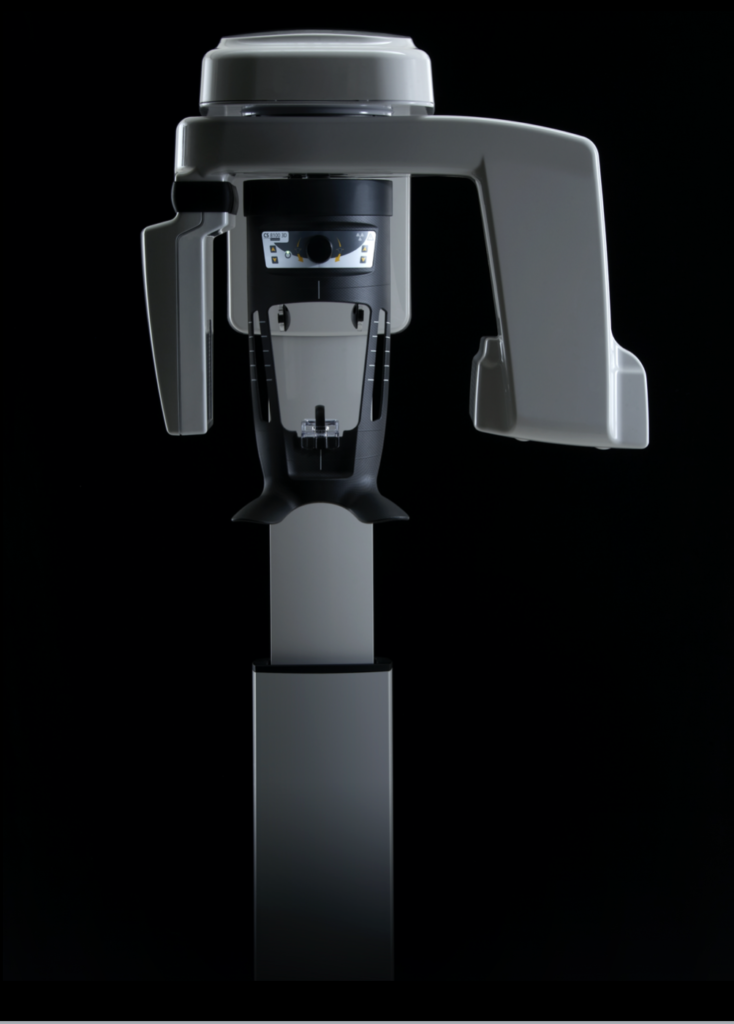
Cone beam computed tomography (CBCT) is a recent advancement in dental imaging and provides a 3D image of the tooth and jaw with high spatial resolution and diagnostic ability.
The dental CBCT does not replace conventional 2D x-rays, but is an additional tool that your dentist or periodontist uses to gain more information, to avoid exploratory surgery, and to perform a ‘virtual surgery’.
A CBCT works by three basic steps: Acquisition, Reconstruction, and Analysis
1. Acquisition
CBCT technology uses the same basic technology of conventional 2D x-rays. That is an x-ray source sends a beam of x-rays through the object of interest and an x-ray detector collects them on the other side.
As the x-ray beam travels through the object of interest (i.e., our teeth), some x-rays pass through the less dense material and others are absorbed by the dense tissue.
For example; If your tooth has a crack in it, the x-ray beam will be absorbed by the tooth, but will pass right through the empty space of the crack and hit the detector. This paints a picture on the detector of a solid tooth with an empty space through it. Voila! Your dentist or periodontist can now confirm a crack in 3D, something they would have missed with the low resolution of a 2D x-ray.
The major difference in the acquisition stage between a 2D and 3D image is the sheer number of ‘pictures’ taken. With 2D technology, only one photo is captured at a single angle. With 3D technology, many many photos are captured as the x-ray source and detector move around the patients head.
2. Reconstruction
After all of the individual images are acquired, sophisticated software reconstruct the images to re-create the 3D volume.
Like a Rubik’s cube, the individual images are stacked on top of one another in the sequence they were taken in to build a 3D cube.
3. Analysis and Interpretation
The final stage involves the analysis and interpretation of the images captured. A great benefit to CBCT technology is the ability to manipulate, maneuver and rotate the 3D volume to scroll through the “stack” of images created.
This drastically improves the ability of your dentist or periodontist to diagnose and plan treatment accordingly.
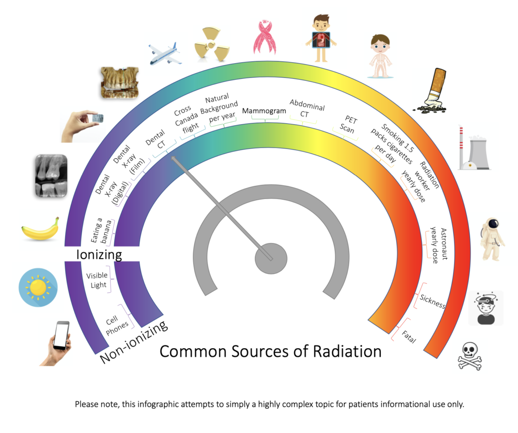
A question that we are asked all the time is “How much radiation is this going to expose me to?”
This is a highly complex question, with so many factors influencing the answer for each individual patient. The simple answer is, not more than three dental film x-rays but the information we receive from a single CBCT is exponentially more!
There are many factors and settings in the acquisition stage of the CBCT scan that can be manipulated by the dentist or periodontist to limit the radiation exposure to the patient. To stay informed, always ask your prescribing dentist or periodontist how they plan to limit you to the lowest possible radiation exposure.


